Multimodal optical imaging

Defects on surfaces of optical materials limit the performance of high energy and high peak power laser systems.
We use the latest high sensitivity and high-resolution optical characterization tools to understand laser-materials interactions.
Photoluminescence lifetime imaging

Optical materials undergo laser-induced damage due to defects created by fracture and contamination.
Imaging these defects in 3D, and including photoluminescence lifetime information allows PLS scientists to understand mechanisms of defect creation and mitigation strategies.
High-resolution, high-sensitivity transmission loss imaging (down to 0.1%) is used to measure the effects of exposures to many low and high fluence laser pulses.
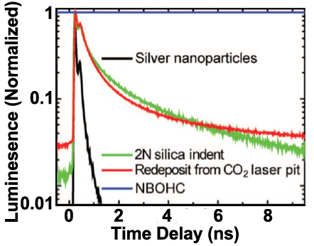
Photoluminescence lifetime decays reveal fast lifetimes not associated with known silica point defects. (Applied Physics Letters 94, 151114, 2009)
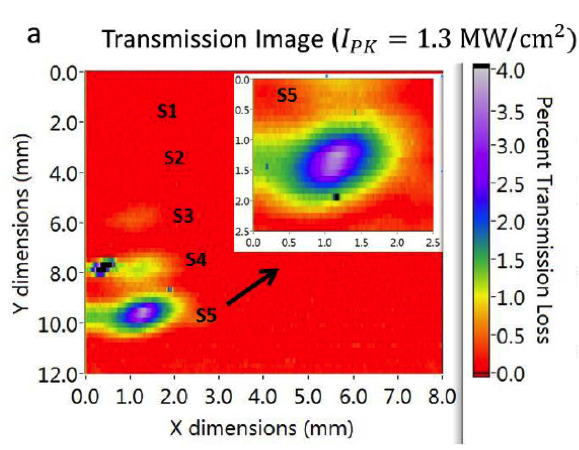
Transmission loss on fused silica surface caused by exposure of 1 billion shots at 0.1 J/cm2 in vacuum. (Ly et al, Optics Express, 23, 4074-4091)